Presentation

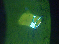
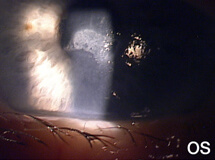
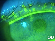
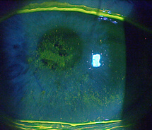
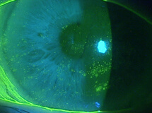
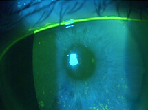
A 78-year-old female was referred to my clinic for evaluation of corneal abrasions and mucopurulent discharge that had persisted for at least 3 to 4 weeks. Her chief complaint was that her eyes felt like they were “full of sand,” and she was unable to see well. She also described both eyes as burning, painful, itchy, red, and sensitive to light. She was an avid reader but had not been able to read for over a year. At the time of her referral, she was using artificial tears every 2 hours and erythromycin ointment BID.

The patient had a longstanding history of moderate to severe dry eye, for which she had been offered and tried numerous treatments over the years, including artificial tears, corticosteroids, topical cyclosporine, punctal occlusion, and autologous serum. She had had cataract surgery about 8 years prior, and she stated that she had been given antiviral therapy for unilateral “red eye” about 15 years ago. Her medical history was significant for diabetes.

The patient’s symptoms of foreign body sensation, blurry vision, eye pain, and photophobia appear to be consistent with corneal abrasion, but she lacked a definite history of ocular injury, the most common cause of corneal abrasion.1 The discharge she reported caused me to suspect an infection.

  1. Fraser S. Corneal abrasion. Clin Ophthalmol. 2010;4:387-90.

Based on this patient’s history, what condition(s) do you think may have led to the spontaneous, persistent abrasion?

 
a. Dry eye
Please select again. Yes, persistent corneal epithelial defects can develop secondary to some primary ocular surface disorders including dry eye.1 What else could be the cause of her condition?
  1. Poon AC, Geerling G, Dart JK, et al. Autologous serum eyedrops for dry eyes and epithelial defects: clinical and in vitro toxicity studies. Br J Ophthalmol. 2001;85:1188-97.
 
b. Prior viral keratopathy
Please select again. Yes. Herpes simplex viruses have an affinity for the sensory ganglion cells and the potential to cause corneal sensory impairment; it is not uncommon for patients with prior herpes simplex keratitis to develop nonhealing epithelial defects and ulcers without actively replicating virus. 1 What else could be the cause of her condition?
  1. Sacchetti M, Lambiase A. Diagnosis and management of neurotrophic keratitis. Clin Ophthalmol. 2014;8:571-9.
 
c. Diabetes
Please select again. Yes. Diabetic patients are at higher risk of developing corneal complications including persistent epithelial defects.1 What else could be the cause of her condition?
  1. Lutty GA. Effects of diabetes on the eye. Invest Ophthalmol Vis Sci. 2013;54(14):81-7.
 
d. All of the above
Yes. The patient had more than one condition—ocular and systemic—that could underlie her corneal lesion: advanced dry eye and herpes simplex keratitis can both induce persistent epithelial defects;1, 2 and keratopathy, including recurrent corneal erosions and persistent epithelial defects, is a well-established ocular complication of diabetes.3
  1. Poon AC , Geerling G, Dart JK, et al. Autologous serum eyedrops for dry eyes and epithelial defects: clinical and in vitro toxicity studies. Br J Ophthalmol. 2001;85:1188-97.
  2. Sacchetti M, Lambiase A. Diagnosis and management of neurotrophic keratitis. Clin Ophthalmol. 2014;8:571-9.
  3. Lutty GA. Effects of diabetes on the eye. Invest Ophthalmol Vis Sci. 2013;54(14):81-7.

Examination

The patient’s uncorrected visual acuity was 20/400 OD and hand motion OS. Her vision improved with a pinhole to 20/200 OD, but not in the left eye.

Slit lamp examination revealed diffuse, grade 4 superficial punctuate keratitis in both eyes and a 1.5 x 2.5-mm central epithelial defect with Descemet’s folds and corneal edema in the left eye. There were no significant signs of ocular inflammation or stromal involvement. Both eyes had trace conjunctival injection and grade 2 meibomian gland dysfunction (MGD). Her tear meniscus height measured 0.2 microns. Tear osmolarity was 326 mOsm/L OD and 339 mOsm/L OS.

After ocular surface evaluation, I applied a cotton wisp strand to the cornea. In this test, what condition do you think I sought to pinpoint?

 
a. Corneal hypoesthesia/anesthesia
Yes, innervation is important for maintaining the normal structure and function of the cornea, and reduced corneal sensitivity can be associated with epithelial defects in many corneal diseases.1 With her history of viral infection and diabetes and the nonhealing nature of the epithelial defect, I suspected impairment of corneal sensation in this patient.
  1. Aragona P, Stilo A, Ferreri F, et al. Effects of the topical treatment with NSAIDs on corneal sensitivity and ocular surface of Sjögren's syndrome patients. Eye (Lond). 2005;19(5):535-9.
 
b. Decreased tearing reflex
Please select again. No, I did not consider tearing reflex pertinent to this patient’s condition.
 
c. Loose adhesion of the epithelium
Please select again. This was not my consideration.
 
d. Incomplete blink
Please select again. This was not my consideration although it is an important finding to evaluate.

Diagnosis

The patient demonstrated significant reduction of corneal sensitivity in both eyes, with sensitivity in the left eye reduced to near zero.

Based on the patient’s history and examination findings, what do you think my principal diagnosis was?

 
a. Chronic epithelial defect associated with her severe dry eye disease
Please select again. There is no doubt that this patient had severe dry eye—she had a history of dry eye, prominent MGD, scant tear meniscus height, and tear hyperosmolarity, but I did not consider dry eye to be the primary cause of her corneal lesion.
 
b. Herpetic epithelial keratitis
Please select again. This was not my diagnosis. Characterized by a central location and Descemet’s membrane folds and stromal swelling, this patient’s corneal lesion lacked the distinctive features of an active herpetic viral infection, such as dendritic or geographic ulceration and significant stromal inflammation. Additionally, herpetic epithelial disease rarely persists the way this patient’s had. It is typically self-limited and will resolve without intervention.
 
c. Neurotrophic keratitis
Yes. Poor epithelial healing and loss of corneal sensitivity are characteristic features of neurotrophic keratopathy.1 In this patient, the diagnosis of neurotrophic keratopathy is further supported by history of viral infection (potentially herpes simplex keratopathy associated with corneal sensory impairment) and the appearance of the epithelial defect.1 According to the classification system of neurotrophic keratitis based on severity, her condition was more advanced in the left eye (stage II, characterized by persistent epithelial defect) than the right (stage I, characterized by superficial punctate keratitis).1 This implies that previous herpetic keratopathy likely played a more important role in the development of the epithelial defect, though diabetes might have predisposed both eyes to neurotrophic keratopathy.
  1. Sacchetti M, Lambiase A. Diagnosis and management of neurotrophic keratitis. Clin Ophthalmol. 2014;8:571-9.
 
d. Toxic ulcerative keratoconjunctivitis

Restoring Corneal Integrity


The patient received an amniotic ring in the left eye and extended-duration punctal plugs in both eyes. She continued with the erythromycin ointment qhs, a lubricating drop with 2% polyvinyl pyrrolidone and polyvinyl alcohol, and preservative-free artificial tear. Four days later the amniotic membrane ring was removed. The epithelial defect had shrunk to 0.5 x 1 mm. The patient stated that her left eye felt better but still had some discomfort.

What topical medication do you think I prescribed for this patient after removal of the amniotic ring device?

 
a. A corticosteroid
Please select again. Corticosteroids are not indicated in this case as the patient demonstrated no marked inflammatory reaction. In fact, corticosteroids should be avoided in patients with neurotrophic keratitis because they may increase collagenase activity and stromal melting.1
  1. Sacchetti M, Lambiase A. Diagnosis and management of neurotrophic keratitis. Clin Ophthalmol. 2014;8:571-9.
 
b. A non-steroidal anti-inflammatory drug (NSAID)
Please select again. It is recommended that topical NSAIDs be avoided in the treatment of neurotrophic keratitis patients because of their potential to inhibit epithelial healing and reduce corneal sensitivity.1,2
  1. Sacchetti M, Lambiase A. Diagnosis and management of neurotrophic keratitis. Clin Ophthalmol. 2014;8:571-9.
  2. Aragona P, Stilo A, Ferreri F, et al. Effects of the topical treatment with NSAIDs on corneal sensitivity and ocular surface of Sjögren's syndrome patients. Eye (Lond). 2005;19(5):535-9.
 
c. An antibiotic
Yes, I continued her prescription of an antibiotic ointment until the epithelial defect and her infection healed. For this patient, I preferred use of an ointment dosage form.
 
d. None of the above
Please select again. I prescribed one of the listed agents for the patient.

Improving and Protecting the Ocular Surface

The abrasion healed within 3 days after removal of the amniotic ring. In the next 3 months, the patient’s vision improved significantly, to 20/70 OD and 20/100 OS, respectively.

The patient was continued on a regimen of gel artificial tears and preservative-free artificial tears. However, about 7 months after her first episode of corneal abrasion, a 2- x 2.5-mm central epithelial defect recurred in the left eye with reduced vision, leaving her 20/200 OD and 20/400 OS. After insertion of a bandage lens and then another amniotic ring, the epithelial defect healed in 2 weeks and the vision of the left eye improved to 20/70.

She then continued on the preservative-free artificial tear regimen and topical cyclosporine. After 5 months, she was still unable to read (she was 20/200 OD and 20/100 OS), and moderate to severe superficial punctuate keratopathy continued to exist in both eyes. By then she had begun to have problems putting drops in the eyes so often.

I suggested LACRISERT® (hydroxypropyl cellulose ophthalmic insert) to the patient. Why do you think I suggested LACRISERT for this patient?

 
a. Once-daily dosing
Please select again. Yes, once-daily dosing contributed to my suggestion of LACRISERT®. For most patients, once daily use of the ophthalmic insert is sufficient.1 Some patients may require twice-daily dosing for optimal results. This is not the only factor I was considering regarding use of LACRISERT for this patient.
  1. Lacrisert [package insert] Bridgewater, NJ: Valeant Pharmaceuticals International, Inc.; 2014.
 
b. Continuous lubrication and protection
Please select again. Yes, lubrication and protection of the corneal surface is important in improving symptoms and signs of this patient’s moderate to severe dry eye. Containing 5 mg of hydroxypropyl cellulose that is released in a sustained fashion over 14 to 18 hours, the LACRISERT® insert acts to lubricate and protect the corneal epithelium with once-daily dosing.1 Some patients may require twice-daily use for optimal results. This is not the only factor I was considering regarding use of LACRISERT for this patient.
  1. Lacrisert [package insert] Bridgewater, NJ: Valeant Pharmaceuticals International, Inc.; 2014.
 
c. Free of preservatives
Please select again. Yes, discontinuation or avoidance of preservatives in topical medications, which may be detrimental to the ocular surface epithelium, can be an important treatment strategy in patients with neurotrophic keratitis.1 What other benefits may LACRISERT® have?
  1. Baudouin C, Labbé A, Liang H, et al. Preservatives in eyedrops: the good, the bad and the ugly. Prog Retin Eye Res. 2010; 29(4):312-334.
 
d. All of the above
Yes, LACRISERT® makes an appropriate therapy for this patient in many ways. Unlike artificial tears, the insert can be dosed once daily; some patients may require twice-daily use for optimal results. The insert contains no preservatives or other ingredients than hydroxypropyl cellulose. Importantly, LACRISERT can provide lubrication and protection of the corneal surface — a key to achieving the treatment goal at this stage: preventing the recurrence of epithelial breakdown.

Follow-up

The patient was willing to try LACRISERT® (hydroxypropyl cellulose ophthalmic insert). After being counseled and given a training session by a technician, she began to use LACRISERT once daily in the morning. Since then, the patient had continued to improve.

Four months after she began the LACRISERT therapy, the patient felt that her eyes were doing much better. Her vision improved to 20/60-1 OD and 20/60 OS; superficial punctate keratopathy had decreased to grade 2 in the right eye and grade 2- in the left.

Given the improvement in her symptoms and corneal conditions, a manifest refraction was performed. She received a pair of new glasses, which corrected her vision to 20/50 OD and 20/40-2 OS. She was extremely pleased with her glasses and could now read again.

Congratulations!You’ve completed Case Study 2.

Jack L. Schaeffer, OD, FAAO

Paul Karpecki, OD, FAAO Works in Corneal Services and is clinical research director at the Koffler Vision Group in Lexington, KY. He is a consultant for Bausch + Lomb and Bio-Tissue Inc.

Indications and Usage

LACRISERT® (hydroxypropyl cellulose ophthalmic insert) is indicated in patients with moderate to severe dry eye syndromes, including keratoconjunctivitis sicca. LACRISERT® is indicated especially in patients who remain symptomatic after an adequate trial of therapy with artificial tear solutions. LACRISERT® is also indicated for patients with exposure keratitis, decreased corneal sensitivity, and recurrent corneal erosions.

Important Safety Information

  • LACRISERT® (hydroxypropyl cellulose ophthalmic insert) is contraindicated in patients who are hypersensitive to hydroxypropyl cellulose.
  • Instructions for inserting and removing LACRISERT® should be carefully followed.
  • If improperly placed, LACRISERT® may result in corneal abrasion. Because LACRISERT® may cause transient blurred vision, patients should be instructed to exercise caution when driving or operating machinery.
  • The following adverse reactions have been reported, but were in most instances mild and temporary: transient blurring of vision, ocular discomfort or irritation, matting or stickiness of eyelashes, photophobia, hypersensitivity, eyelid edema, and hyperemia.

To report suspected adverse reactions, contact Bausch & Lomb Incorporated at 1-800-321-4576 or FDA at 1-800-FDA-1088 or FDA.gov/medwatch.

Click here for full Prescribing Information

Indications and Usage

LACRISERT® (hydroxypropyl cellulose ophthalmic insert) is indicated in patients with moderate to severe dry eye syndromes, including keratoconjunctivitis sicca. LACRISERT® is indicated especially in patients who remain symptomatic after an adequate trial of therapy with artificial tear solutions.

Important Safety Information

  • LACRISERT® (hydroxypropyl cellulose ophthalmic insert) is contraindicated in patients who are hypersensitive to hydroxypropyl cellulose.
  • Instructions for inserting and removing LACRISERT® should be carefully followed.

Important Safety Information

LACRISERT® (hydroxypropyl cellulose ophthalmic insert) is contraindicated in patients who are hypersensitive to hydroxypropyl cellulose.